



| HEADLINE |

|

MIT researchers develop injectable ‘mini livers’ to support damaged organs
Published : Monday, 7 September, 2026 at 1:18 PM
Observer Online Desk


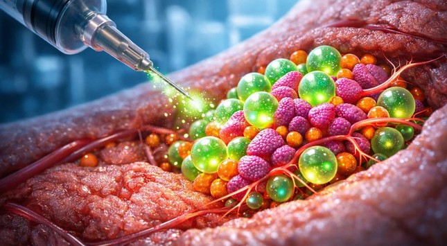
Scientists at the Massachusetts Institute of Technology (MIT) have developed an injectable “mini liver” system that could one day help restore some essential functions of a damaged liver without requiring a surgical transplant.
In a study involving mice, researchers found that injected liver cells remained alive and functional for at least eight weeks, continuing to produce many of the enzymes and proteins normally made by the liver.
The study, published in Cell Biomaterials, was led by MIT postdoctoral researcher Vardhman Kumar, with Sangeeta Bhatia, a professor at MIT and member of its Koch Institute for Integrative Cancer Research, as senior author.
The liver performs around 500 essential functions in the body, including regulating blood clotting, removing bacteria from the bloodstream and processing medications. Many of these functions are carried out by specialized cells called hepatocytes.
For more than a decade, Bhatia's laboratory has explored ways to restore hepatocyte function without requiring patients to undergo liver transplantation. Earlier approaches involved placing liver cells inside biomaterials such as hydrogels, but those materials had to be surgically implanted.
The new technique uses an injectable mixture containing hepatocytes and tiny hydrogel microspheres. The spheres help keep the liver cells together and promote connections with nearby blood vessels.
When densely packed, the microspheres allow the mixture to flow like a liquid through a syringe. After injection, however, the material regains a more solid structure, creating a stable environment for the transplanted cells.
The researchers also included fibroblast cells, which help support hepatocyte survival and stimulate the growth of new blood vessels around the graft.
Using an ultrasound-guided syringe, the team injected the mixture into fatty tissue in the abdomen of mice. Ultrasound can subsequently be used to monitor the graft and determine whether it remains stable.
Following injection, the liver cells formed a compact structure and gradually developed connections with newly formed blood vessels. Those vessels supplied nutrients and helped the hepatocytes remain alive and functional.
The cells survived throughout the eight-week experiment and continued releasing specialized proteins into the animals' bloodstream, suggesting that the grafts could potentially provide longer-term liver support.
Researchers said the mini livers do not necessarily have to be located next to the patient's liver. They could potentially be placed in other areas with adequate space and blood supply, including the spleen or areas near the kidneys.
The technology could eventually serve several purposes, including providing an alternative to surgical transplantation or temporarily supporting patients while they wait for a donor organ.
“If we think they might need another therapy or more grafts, the barriers to do that are much less with this injectable technology than undergoing another surgery,” Kumar said.
The approach would currently likely require patients to take immunosuppressive drugs to prevent rejection of the transplanted cells. Researchers are exploring ways to overcome that limitation, including developing hepatocytes capable of avoiding immune detection and using the hydrogel microspheres to deliver immunosuppressive drugs directly around the graft.
The researchers said further studies will be needed before the technology can be tested in humans.
In a study involving mice, researchers found that injected liver cells remained alive and functional for at least eight weeks, continuing to produce many of the enzymes and proteins normally made by the liver.
The study, published in Cell Biomaterials, was led by MIT postdoctoral researcher Vardhman Kumar, with Sangeeta Bhatia, a professor at MIT and member of its Koch Institute for Integrative Cancer Research, as senior author.
The liver performs around 500 essential functions in the body, including regulating blood clotting, removing bacteria from the bloodstream and processing medications. Many of these functions are carried out by specialized cells called hepatocytes.
For more than a decade, Bhatia's laboratory has explored ways to restore hepatocyte function without requiring patients to undergo liver transplantation. Earlier approaches involved placing liver cells inside biomaterials such as hydrogels, but those materials had to be surgically implanted.
The new technique uses an injectable mixture containing hepatocytes and tiny hydrogel microspheres. The spheres help keep the liver cells together and promote connections with nearby blood vessels.
When densely packed, the microspheres allow the mixture to flow like a liquid through a syringe. After injection, however, the material regains a more solid structure, creating a stable environment for the transplanted cells.
The researchers also included fibroblast cells, which help support hepatocyte survival and stimulate the growth of new blood vessels around the graft.
Using an ultrasound-guided syringe, the team injected the mixture into fatty tissue in the abdomen of mice. Ultrasound can subsequently be used to monitor the graft and determine whether it remains stable.
Following injection, the liver cells formed a compact structure and gradually developed connections with newly formed blood vessels. Those vessels supplied nutrients and helped the hepatocytes remain alive and functional.
The cells survived throughout the eight-week experiment and continued releasing specialized proteins into the animals' bloodstream, suggesting that the grafts could potentially provide longer-term liver support.
Researchers said the mini livers do not necessarily have to be located next to the patient's liver. They could potentially be placed in other areas with adequate space and blood supply, including the spleen or areas near the kidneys.
The technology could eventually serve several purposes, including providing an alternative to surgical transplantation or temporarily supporting patients while they wait for a donor organ.
“If we think they might need another therapy or more grafts, the barriers to do that are much less with this injectable technology than undergoing another surgery,” Kumar said.
The approach would currently likely require patients to take immunosuppressive drugs to prevent rejection of the transplanted cells. Researchers are exploring ways to overcome that limitation, including developing hepatocytes capable of avoiding immune detection and using the hydrogel microspheres to deliver immunosuppressive drugs directly around the graft.
The researchers said further studies will be needed before the technology can be tested in humans.

Loading...
Loading...
Also read





Editor : Iqbal Sobhan Chowdhury
Published by the Editor on behalf of the Observer Ltd. from Globe Printers, 24/A, New Eskaton Road, Ramna, Dhaka.
Editorial, News and Commercial Offices : Aziz Bhaban (2nd floor), 93, Motijheel C/A, Dhaka-1000.
Phone: PABX- 41053001-06; Advertisement: 41053012; 01793317829, 01550707291, E-mail: [email protected], [email protected] Online: email: [email protected] 41053014; 01550707297 Advertisement: 01550707296
Published by the Editor on behalf of the Observer Ltd. from Globe Printers, 24/A, New Eskaton Road, Ramna, Dhaka.
Editorial, News and Commercial Offices : Aziz Bhaban (2nd floor), 93, Motijheel C/A, Dhaka-1000.
Phone: PABX- 41053001-06; Advertisement: 41053012; 01793317829, 01550707291, E-mail: [email protected], [email protected] Online: email: [email protected] 41053014; 01550707297 Advertisement: 01550707296


